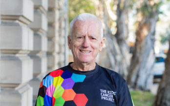
Recipient: Dr Joseph Yuan-Mau Yang
Institute: Murdoch Children’s Research Institute
Funding: $275,962.00
Dr Joseph Yuan-Mou Yang is the lead clinician scientist for the Neuroscience Advanced Clinical Imaging Service, Department of Neurosurgery, at the Royal Children’s Hospital, Melbourne. Dr Yang’s research focuses on developing and implementing advanced neuroimaging techniques to help diagnostics and surgical treatment capacities for children with brain tumours and other surgical amendable neurological conditions. With a medical doctoral background in neurosurgery and a PhD investigating the use of advanced brain nerve fibre tract imaging (tractography) in paediatric epilepsy surgery, Dr Yang also holds honorary positions at the co-located Murdoch Children’s Research Institute, and Melbourne University.
Motivation
My clinical experience in neurosurgery training and practice over the last 15 years has inspired me to help advance brain tumour research in children. Over the past decade, I have witnessed the tremendous progress made in tumour genetic research which is changing the landscape of neuro-oncology practice. The findings that both a multidisciplinary approach to brain tumour care and a close engagement between clinicians and researchers is the key to clinical translation success.
In 2012, I undertook a neuroimaging PhD that investigated the use of advanced brain nerve fibre tract imaging technique (a.k.a. tractography) to help surgical planning and image guidance to improve the precision of brain surgery performed in children with drug-resistant epilepsy and brain tumours. In contrast to the progress being made in cancer molecular biology and genetics, the translation of advanced neuroimaging techniques into the clinical neurosurgery realm to help improve brain surgery safety profile was lacking.
More worryingly, the commercial-based and widely used tractography technique assisting surgery was based on outdated technology, potentially harming the patient by providing inaccurate information to the operating neurosurgeon. Despite the thriving research, advanced tractography has not reached the clinics. My clinical experience and success in implementing advanced tractography over the last decade at the Royal Children’s Hospital has made me even more determined to help bridge this clinical research gap, and to undertake the challenge of developing advanced tractography techniques, making access to precision surgical care more equitable for other neurosurgery centres, both locally and globally.
AI-guided brain imaging
Brain tumours in children are abnormal growths of cells located somewhere on or in the brain. These tumours are either benign or malignant and arise from different types of brain cells occurring in various parts of the brain, leading to a wide range of symptoms and outcomes. Brain tumours are the second most common type of cancer in children (behind leukaemia). They are among the leading causes of childhood disease-related death in Australia and worldwide.
Brain surgery is the mainstay treatment that reduces brain tumour burden and prolongs survival. However, surgery performed near functional brain regions and the interconnecting nerve fibre tracts has a high morbidity risk. Advanced MRI-based nerve fibre tract imaging (tractography) can greatly assist with post-surgical functional preservation by avoiding surgical tract injuries. Existing commercial surgical tractography software used by most neurosurgical centres worldwide is based on outdated techniques that provide anatomically inaccurate tractography images, leading to a great risk of surgical tract injuries.
My project aims to develop and implement an automated surgical tractography technique leveraging my expert knowledge and artificial intelligence (AI). This project will develop an AI-based technique to reconstruct the three most important brain nerve fibre tract images used in planning childhood brain tumour surgery, each corresponding to the carrying out of motor, language, and visual functions. The intended outcomes of this project will be to accelerate the complex image reconstruction and analysis needed for neurosurgical planning, enable uptake of our robust neuroimaging pipeline outside of our dedicated clinical service, and provide robust, reliable, and informative directions for critical neurosurgical operations, in both acute emergency and elective surgical settings.
In the past ten years, the advent of powerful genomic and drug discovery technologies has contributed to improved outcomes, yet brain surgery remains the mainstay treatment to reduce tumour burden and prolong survival. The accuracy of brain surgery is paramount, especially for tumours located near brain regions carrying important bodily functions, such as movement, language, and vision. Surgical injuries to these critical functional brain regions, including the underlying connecting nerve fibres, can result in devastating, permanent functional damage adversely impacting the quality of tumour survivorship.
The prognosis and outcomes for children with brain tumours can vary widely depending on factors such as tumour location, genetics, and response to both surgical and adjuvant treatment. Research and advancements in treatment continue to improve outcomes for children with brain tumours, but comprehensive, multidisciplinary care remains critical for the best possible results. Close collaboration between paediatric oncologists, neurosurgeons, radiation oncologists, and other specialists is essential to optimise treatment plans and outcomes.
Advancing brain imaging technology
The primary focus of my research project is to develop and directly translate an innovative tractography technique to help make brain surgery safer for children. With a greater likelihood of complete tumour removal, affected children and adolescents and their families, will directly benefit via a reduction in the severity of post-surgical functional deficits, maximising their quality of life. My project will address the technical shortcomings of existing commercial tractography solutions, leading to more accurate mapping of brain nerve fibre tract images adjacent to the tumour. The artificial intelligence basis of the tractography technique would ensure all children with brain tumours undergoing brain surgeries in both acute emergency and elective settings, have equal access to and benefit from the availability of this advanced brain imaging technology.
My research will also contribute to a more comprehensive presurgical evaluation of surgical risks; and improved mapping of safe surgical strategy during presurgical planning. Safe surgeries can be offered to individuals who were previously considered inoperable due to perceived high surgical risks resulting from inadequate presurgical evaluation. The ability to deliver safe and personalised precision brain surgery in a timely manner, informed by the proposed advanced tractography technique, can contribute significantly towards maximising the quality of life for all brain tumour survivors and their families.
Closing a clinical gap
This research addresses a clear and critical clinical research gap concerning the tractography technology used in routine neurosurgery practice. In particular, the gap highlights the lack of implementing advanced tractography techniques in the clinical realm, and the inability to provide advanced tractography images to acute brain tumour surgery settings due to the relatively long data processing time that’s incompatible with the surgical acuity.
Existing commercial surgical tractography software, used by most neurosurgical centres worldwide, is based on outdated techniques that provide anatomically inaccurate tractography images, leading to a great risk of surgical tract injuries. Similarly, current artificial intelligence-driven tractography methods available in the research domain are incredibly useful for tracking healthy brain structures but are not suitable for children with brain tumours given the large changes to brain structure in small-to-large tumours.
My research is important to help close this clinical research gap and can also help reduce the currently high barriers to using advanced tractography for brain tumour surgery, making access to precision surgical care more equitable across multiple neurosurgery centres, both locally and worldwide.
Col Reynolds Fellowship
I feel extremely privileged and very proud! It’s truly an honour to be an inaugural recipient of the Col Reynolds Fellowship. This Fellowship adds to my ongoing effort of introducing cutting-edge neuroimaging techniques to help improve the safety of brain surgeries conducted in children with brain tumours and other surgical neurological conditions. This Fellowship will help advance my career significantly, to be seen both as a pioneer and a world-leading expert, implementing, and realising the values of advanced neuroimaging techniques to clinical neurosurgery practice. It will provide me with the opportunity to engage with neurosurgeons and diffusion MRI tractography research experts both locally and internationally. Multi-centre collaboration and data pooling are key elements of seeing the translation success and the diversity of clinical MRI datatypes from different institutions will improve the likelihood of widespread clinical uptake of this research output.
With the advent of brain tumour genetics and associated drug therapies over the last decade, we have entered a ‘golden’ era of brain tumour therapy where we have begun to see improvements in survival and importantly, the quality of survivorship. I believe the outcome of this Fellowship will contribute significantly towards advancing surgical care for children with brain tumours, and complement the advancements made in brain tumour biology and genetics. Such a multidisciplinary and multi-faceted approach to clinical translation will ensure these children receive the best possible care, with the intention to deliver them with the best possible clinical outcomes.
Thank you, donors!
Please accept my heartfelt gratitude for this generous gift. This Fellowship would not be possible without your generous donation. Together, this opportunity will put us in a great position to help make life-changing improvements to the way brain surgery is performed in children with brain tumours. I am looking forward to starting the work, and to sharing with you the emerging impact of this work on neurosurgery practice, both in Australia and worldwide.
Extra thanks
I want to thank The Kid’s Cancer Project again for this Fellowship award. I want to thank my surgical and career mentor, Ms Wirginia Maixner and my Head of Department, Ms Alison Wray, for their mentorship and guidance. I want to thank my team members, Dr Sila Genc and Dr Bonnie Alexander, my supervisors, Dr Gareth Ball and A/Prof Richard Beare, the Murdoch Children’s Research Institute grants office, and my internal and external collaborators for their unconditioned support for this Fellowship application.
Find out more about the Col Reynolds Fellowship
With an investment of over $7.6 million, The Kids’ Cancer Project is ensuring that some of the best and brightest young researchers in Australia can further their careers and most importantly, their impact on childhood cancer research.


Read more from past recipients
From a field of outstanding candidates across Australia, The Kids’ Cancer Project has funded the next generation of childhood cancer researchers. Their science-backed research is sure to deliver breakthroughs across a range of areas relating to childhood cancer.
